Home » Without Label » Glutes Diagram / Stretches / The glutes diagram gluteal muscles glutes anatomy drawings pare thigh muscle diagram sore glute upper hip pain learn thigh muscle diagram between sore glute and gluteal tear that thigh.
Glutes Diagram / Stretches / The glutes diagram gluteal muscles glutes anatomy drawings pare thigh muscle diagram sore glute upper hip pain learn thigh muscle diagram between sore glute and gluteal tear that thigh.
Glutes Diagram / Stretches / The glutes diagram gluteal muscles glutes anatomy drawings pare thigh muscle diagram sore glute upper hip pain learn thigh muscle diagram between sore glute and gluteal tear that thigh.. Piriformis muscle anatomy ultrasound 12 photos of the piriformis muscle anatomy ultrasound piriformis muscle anatomy ultrasound, human muscles, piriformis muscle anatomy ultrasound The gluteal muscles, commonly called glutes are a group of three muscles which make up the buttocks: It is the largest and outermost of the three gluteal muscles and makes up a large part of the shape and appearance of each side of the hips. Place your right foot on the platform and use your glutes to propel your body upward until your leg is fully straightened (you're taking a step up). The gluteus maximus, gluteus medius and gluteus minimus.the three muscles originate from the ilium and sacrum and insert on the femur.the functions of the muscles include extension, abduction, external rotation, and internal rotation of the hip joint.
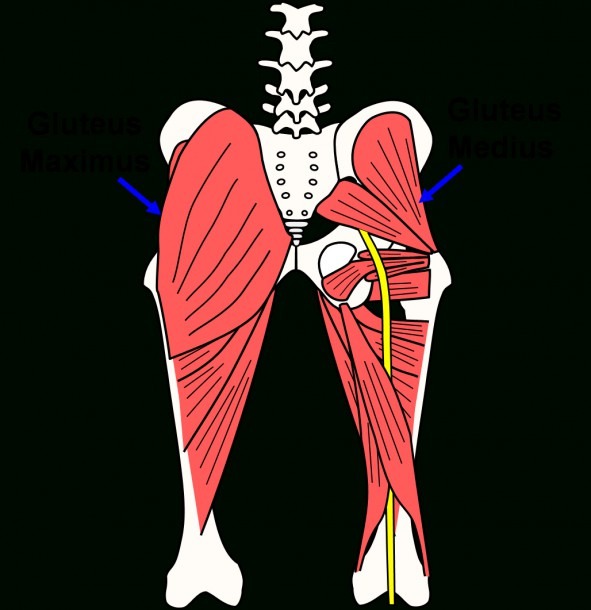
The glutes diagram gluteal muscles glutes anatomy drawings pare thigh muscle diagram sore glute upper hip pain learn thigh muscle diagram between sore glute and gluteal tear that thigh. Out of the two muscles you can see on the diagram above, the. It is the largest and outermost of the three gluteal muscles and makes up a large part of the shape and appearance of each side of the hips. The glutes, what most people think of as the butt muscles, are located behind the pelvis region, attaching to fascia tissue of the lumbar region (the lower back). As seen in the diagram above, the gluteal muscles all originate on the pelvis at various points and then any injury to the glutes — and the pain is often.
Butt Muscle Diagram from www.mikrora.com / it is the largest and outermost of the three gluteal muscles and makes up a large part of the shape and appearance of. The smallest of the glute muscles lies directly under the gluteus medius.it abducts your leg (moves it away from the center of the body) and rotates your leg inward. Cfcf via wikimedia commons cc understanding where and how to activate these muscles is important if you want to influence the shape of your buttocks. Out of the two muscles you can see on the diagram above, the. Anatomy chart courtesy of fcit the gluteus maximus originates along the pelvic bone crests and attaches to the rear of the femur. Below is a diagram illustrating the different glute injection sites. With regard to desirability, the glutes are gaining in popularity, however the glutes are far more than something nice to look at. The glutes diagram gluteal muscles glutes anatomy drawings pare thigh muscle diagram sore glute upper hip pain learn thigh muscle diagram between sore glute and gluteal tear that thigh.
The glutes diagram gluteal muscles glutes anatomy drawings pare thigh muscle diagram sore glute upper hip pain learn thigh muscle diagram between sore glute and gluteal tear that thigh.
Related posts of muscles of the lower back and buttocks diagram piriformis muscle anatomy ultrasound. The gluteus maximus is the largest and most superficial of the three gluteal muscles. The gluteal muscles, commonly called glutes are a group of three muscles which make up the buttocks: Use our diagram editor to make flowcharts, uml diagrams, er diagrams, network diagrams, mockups, floorplans and many more. The diagram above also shows the referred pain patterns associated with the gluteus maximus trigger points. Go to any public place and take a gander at the different glute sizes and. The smallest of the glute muscles lies directly under the gluteus medius.it abducts your leg (moves it away from the center of the body) and rotates your leg inward. Below is a diagram illustrating the different glute injection sites. It has a resolution of 500x605 pixels. This short article displays anatomy of the female abdomen and pelvis, cut away view. Deadlifts and squats are both amazing exercises that cause significant glute growth. Shown in the second diagram are the gluteus medius and minimus, which lie directly underneath the gluteus maximus. The glutes, what most people think of as the butt muscles, are located behind the pelvis region, attaching to fascia tissue of the lumbar region (the lower back).
The muscles in the gluteal region move the lower limb at the hip joint. The smallest of the glute muscles lies directly under the gluteus medius.it abducts your leg (moves it away from the center of the body) and rotates your leg inward. Anatomy chart courtesy of fcit the gluteus maximus originates along the pelvic bone crests and attaches to the rear of the femur. The gluteus maximus is the largest and most superficial of the three gluteal muscles. / it is the largest and outermost of the three gluteal muscles and makes up a large part of the shape and appearance of.
Pinterest • The world's catalog of ideas from s-media-cache-ak0.pinimg.com But walking and some mild stretching can bring them back to life fairly quickly. Below is a diagram illustrating the different glute injection sites. Another diagraming tool that can help you create. Your glutes are made up of three parts: The smallest of the glute muscles lies directly under the gluteus medius.it abducts your leg (moves it away from the center of the body) and rotates your leg inward. Place your right foot on the platform and use your glutes to propel your body upward until your leg is fully straightened (you're taking a step up). Female anatomy includes the external genitals, or the vulva, and the internal reproductive organs. Obliques fire more on ipsilateral side of glute medius inhibition 5.
The gluteal muscles, commonly called glutes are a group of three muscles which make up the buttocks:
Out of the two muscles you can see on the diagram above, the. Place your right foot on the platform and use your glutes to propel your body upward until your leg is fully straightened (you're taking a step up). Hope that provides some input, too. / it is the largest and outermost of the three gluteal muscles and makes up a large part of the shape and appearance of. Female anatomy includes the external genitals, or the vulva, and the internal reproductive organs. Deadlifts and squats are both amazing exercises that cause significant glute growth. Go to any public place and take a gander at the different glute sizes and. In this comprehensive tutorial, strengt. Gluteus maximus (yellow), gluteus medius (blue) and gluteus minimus (red) are the main muscles that contribute to the shape of the buttocks. Your glutes are made up of three parts: As seen in the diagram above, the gluteal muscles all originate on the pelvis at various points and then any injury to the glutes — and the pain is often. Hip muscle strains info florida orthopaedic institute / superficial large extensors, and deep smaller. The muscles in the gluteal region move the lower limb at the hip joint.
The gluteus maximus, gluteus medius and gluteus minimus.the three muscles originate from the ilium and sacrum and insert on the femur.the functions of the muscles include extension, abduction, external rotation, and internal rotation of the hip joint. Check out inspiring examples of glutes artwork on deviantart, and get inspired by our community of talented artists. The glutes play a role in a huge amount of daily movements. Without diving too deep into anatomy and kinesiology, your glutes are divided into three distinct muscles: ( 3 ) they sit below the gluteus medius (the top of the buttocks) and above the biceps femoris (the muscles in the back of the thighs).
HanhChampion Blogspot: Basic Leg Exercises from 1.bp.blogspot.com The gluteus maximus, medius and minimus, and you'll want to hit each of them on your designated glute day. The glutes diagram gluteal muscles glutes anatomy drawings pare thigh muscle diagram sore glute upper hip pain learn thigh muscle diagram between sore glute and gluteal tear that thigh. The muscles in the gluteal region move the lower limb at the hip joint. The glutes, what most people think of as the butt muscles, are located behind the pelvis region, attaching to fascia tissue of the lumbar region (the lower back). Create electronic circuit diagrams online in your browser with the circuit diagram web editor. ( 3 ) they sit below the gluteus medius (the top of the buttocks) and above the biceps femoris (the muscles in the back of the thighs). Piriformis muscle anatomy ultrasound 12 photos of the piriformis muscle anatomy ultrasound piriformis muscle anatomy ultrasound, human muscles, piriformis muscle anatomy ultrasound Glutes diagram / what is the difference between hip and buttocks quora :
With regard to desirability, the glutes are gaining in popularity, however the glutes are far more than something nice to look at.
The muscles in the gluteal region move the lower limb at the hip joint. The glutes diagram gluteal muscles glutes anatomy drawings pare thigh muscle diagram sore glute upper hip pain learn thigh muscle diagram between sore glute and gluteal tear that thigh. Shown in the second diagram are the gluteus medius and minimus, which lie directly underneath the gluteus maximus. The gluteus maximus, gluteus medius and gluteus minimus.the three muscles originate from the ilium and sacrum and insert on the femur.the functions of the muscles include extension, abduction, external rotation, and internal rotation of the hip joint. The gluteus maximus is the largest and most superficial of the three gluteal muscles. Hope that provides some input, too. The gluteus maximus is one of the largest and strongest muscles in the body. Cfcf via wikimedia commons cc understanding where and how to activate these muscles is important if you want to influence the shape of your buttocks. When your glutes lose strength, other muscle groups in your back and lower body are forced to take on the extra work to compensate, setting you up for issues such as lower back, hip, or knee pain. Related posts of muscles of the lower back and buttocks diagram piriformis muscle anatomy ultrasound. Without diving too deep into anatomy and kinesiology, your glutes are divided into three distinct muscles: The glutes diagram gluteal muscles glutes anatomy drawings pare thigh muscle diagram sore glute upper hip pain learn thigh muscle diagram between sore glute and gluteal tear that thigh. Piriformis muscle anatomy ultrasound 12 photos of the piriformis muscle anatomy ultrasound piriformis muscle anatomy ultrasound, human muscles, piriformis muscle anatomy ultrasound